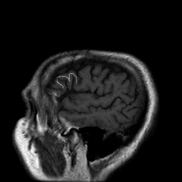


Different scientists have made tremendous progress throughout the 19th and 20th centuries to improve cortical motor mapping. Penfield and Boldrey demonstrated the mapping of motor, sensory, and language cortices by directly stimulating an open cortex in human patients. įritsch and Hitzig (1870) pioneered the discovery of direct electrical cortical stimulation (DCS/DECS) of the animal brain. Research had reported significant beneficial effects from motor mapping, such as the postoperative complication rate dropping from 21% to 13% when adequate neuro-monitoring was employed. The method utilized to obtain such beneficial anatomical and functional knowledge during surgery is called motor mapping. IONM techniques can help identify and find the correct path of the central sulcus (CS) across both hemispheres, while also mapping the cortical homunculus representation at the primary motor area. Knowledge about these alterations caused by tumors allows for safer approaches to surgical resections and guards the patients against potential damage to motor areas as protective actions would be taken. Thus, it is extremely crucial to use methods of locating the functional anatomical regions such as the central sulcus, pre-central gyrus, post-central gyrus, and other functional brain region surrounding the tumor.

Operating on motor regions of the brain becomes even more treacherous when considering how tumor masses may have distorted the anatomical landmarks that surgeons employ for resection surgeries. In contrast, STR calls for the removal of only the necessary parts of the tumor that can potentially alleviate the motor symptoms. It can be challenging to perform in cortical motor surgeries due to the necessity to differentiate between abnormal and healthy tissue. GTR involves the total removal of the tumor. There are two approaches used in tumor resection, gross-total resection (GTR) and subtotal resection (STR). Cortical tumors surrounding the primary motor and premotor areas are likely to cause the symptoms above. The intraoperative neurophysiological monitoring (IONM) of changes to the nervous system caused by surgical manipulations not only helps with lowering postoperative deficits, but it also acts as an alarm system to warn and guide the surgeon. Furthermore, it also demonstrates the need for a safe resection where the odds of patients developing postoperative deficits are reduced. These symptoms justify the need for surgery to alleviate deficits and improve the quality of life for patients if alternative therapeutic effects have failed. Some of the common symptoms caused by motor cortex tumors include hemiparesis or hemiplegia, myopathy, ataxia, gait dysfunction, and spasticity. The patients who have undergone tumor resection surgery can suffer from deficits that can reduce their quality of life. Some other kinds of primary tumors include meningiomas, ependymomas, and many types of lymphomas. A glioma is a tumor that originates from glial cells and is known to be the most common form of malignant primary brain tumor. With primary brain tumors, we can further group them based on the type of cell causing the tumor mass. One such broad division can be made based on tumor malignancy characteristics if the tumor is prone to encroaching on other areas of the body (i.e., malignant) or if it is non-cancerous (i.e., benign). Another method categorizes a tumor as per its origin, which can be termed as primary (i.e., originating in the brain or spinal cord) or secondary (i.e., originating from somewhere else in the body). There are a variety of ways in which one can classify the different kinds of tumors. It resulted in the patient passing away 28 days afterward due to complications from the procedure. The first documented cortical tumor resection was performed by two neurologists A.


 0 kommentar(er)
